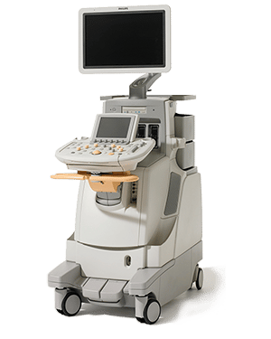
PHILIPS IE33
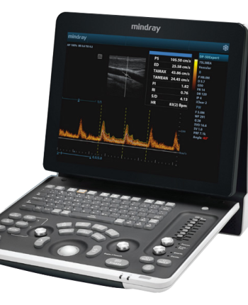
The Philips IE33 premium ultrasound scanner is ideally suited to veterinarians with an interest in cardiology studies and can also be configured for abdominal and soft tissue studies. Clever design and intelligent control are combined to bring you revolutionary clinical performance. The IE33 is configured with veterinary specific applications, coupled with its full cardiac analysis and reporting package, means the IE33 is the perfect system for todays most demanding veterinary hospitals.
An extensive array of available probes means the IE33 can be used over a wide range of cardiac scanning applications from small animals through to equine studies, adding convex and linear probes to the scanner allows it to be used for general soft tissue imaging applications too, making this a true all round scanner.
Philips IE33 Colour Doppler Ultrasound Scanner Probes
Probe compatibility may be system or option specific. Please contact us for further information.
• Phased array probe S5-1 (1-5 mhz)
• Phased array probe S8-3 (3-8 mhz)
• Phased array probe S12-4 (4-12 mhz)
• xMATRIX array probe X3-1 (1-3 mhz)
• xMATRIX array probe X7-2 (2-7 mhz)
• Convex probe C5-1 (1-5 mhz)
• Convex probe C5-2 (2-5 mhz)
• Microconvex probe C8-5 (5-8 mhz)
• Linear probe L 11-3 (3-11 mhz)
• Linear probe L9-3 (3-9 mhz)
• Micro Linear probe L15-7io (7-15 mhz)
• CW probe D2cwc (2 mhz)
• CW probe D5cwc (5 mhz)
• PW probe D2tcd (2 mhz)
• Live 3D TEE probe X7-2t (2-7 mhz)
• Omni TEE probe S7-2 (2-7 mhz)- Categories: Philips, Small Animal, Ultrasound Systems, Veterinary
- Tags: PHILIPS IE33
