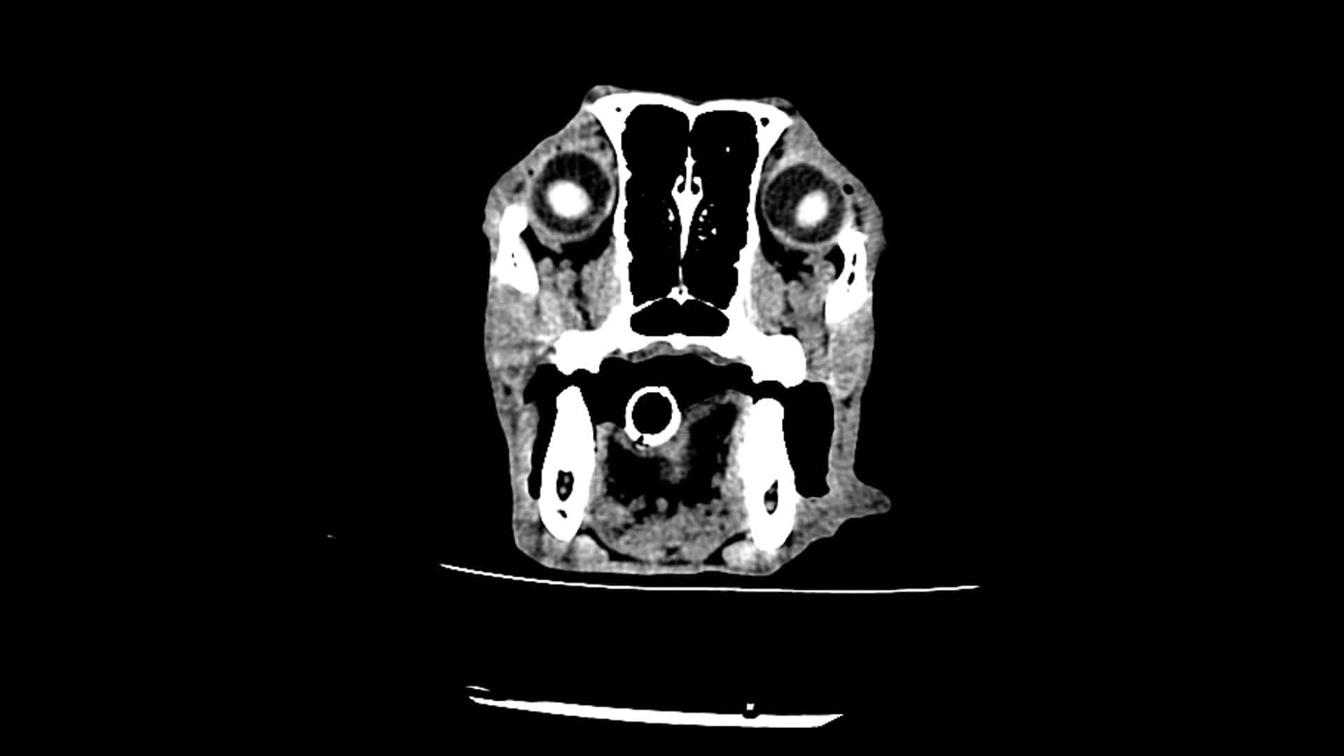
An 8 year old German Shepherd presenting with sudden on set mild seizures. These seizures were well controlled with antiepileptic medication and her neurological examination and haematology and biochemistry were unremarkable.
A CT scan of her brain identified an extra-axial brain mass with strong contrast enhancement compatible with a primary neoplasia originating from the meninges. The primary differential being meningioma especially given the hyperostosis of the parietal bone but other differentials such as histiocytic sarcoma, granular cell tumour or lymphoma existed.
The CT also revealed increased intracranial pressure with moderate transalpine brain herniation and mild transtentorial herniation and incidental small rhinoliths. Whilst these CT findings didn't enable a cure for this patient they greatly assisted the owner with prognosis and our management of her case when seizures increased in frequency and severity.
The CT also assisted our planning for CSF analysis given the evidence of increased intracranial pressure and herniation.
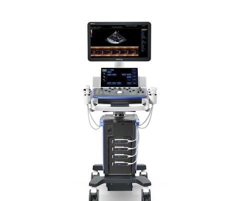
The case study and image was kindly provided by Faye Bethell, of Toll Barn Vets, North Walsham, who uses a 16 slice CT scanner supplied and installed by Probo Medical.